
Page 40 - Spring2020
P. 40
and yet lack the ability to move it into the tissues? Could this increase in supplements is MK-7, not the type we get in
in carboxylation increase the calcification of the placenta or form excess food. The best way to get active and efficiently
osteocalcin, which does cross into the placenta, causing a decrease in assimilated vitamin K is from food. This is
2
the blood supply (thus less oxygen) and explain the findings of small for true of all vitamins. An NIH-funded study
gestational age and other defects in the natto study? involving twenty-seven thousand people over
In 1992, Dr. Hideaki Iioka found that vitamin K MK-4 is transported a six-year period found that “individuals who
2
into the placenta by a carrier protein via an existing transport carrier sys- reported taking dietary supplements had about
tem in the brush border membrane of the human placenta. 18,19,20 Vitamins the same risk of dying as those who got their
A, D and E are also carried in the blood by a carrier protein. Could this nutrients through food. What’s more, the mor-
be the reason that vitamin K MK-4 is often not detected in the blood, tality benefits associated with adequate intake
2
because it is attached to a carrier protein? Little to no research has been of vitamin A, vitamin K, magnesium, zinc, and
done to answer these questions. copper were limited to food consumption.” 23
What we do know is that the traditional sacred foods for preconcep-
tion and pregnancy were foods rich in MK-4, and that traditional weaning TAIL SIZE MATTERS
foods for babies were poultry liver and egg yolk, also great sources of Vitamin K-dependent proteins (VKDP)
vitamin K MK-4. are a group of proteins providing life-giving
2
It’s important to understand that vitamin K MK-4 and long-chain functions for the brain and body. To become bio-
2
MKs are structurally different and are derived from different sources. active they require vitamins K and K MK-4
1
2
Some researchers have suggested a theory of conversion from vitamin as cofactors for the enzyme y-carboxyglutamyl
K MK-7 or other MKs to vitamin K MK-4 via the enzyme UBIAD1, carboxylase (GGCX), which transforms the
1
2
which removes the longer side chains of the K vitamins to produce mena- glutamic acid residues (GLA) in the protein,
dione (K ). K then travels to the liver for detoxification and is somehow promoting calcium-binding and inducing con-
3
3
transported in the blood or lymph by an unexplained carrier to tissues formational changes so that vitamin K can be
where an unknown enzyme(s) adds side chains back to K producing utilized by the tissues. In other words, vitamin
3
vitamin K MK-4. 21 K MK-4 is multifunctional.
2
2
The question is, what happens if K exceeds the rate at which the Once GGCX is activated, vitamin K trans-
3
enzyme can add back the side chains, as when someone is taking K as forms into the epoxide state; then it is recycled to
3
a supplement? Does the excess K cause toxicity and oxidative stress? the quinone and hydroquinone states by vitamin
3
The research is unclear. K epoxide reductase (VKORC1).
What we do know is that K causes disruption or rupture of red blood In 2018, Nolan Chatron and his group used
3
cells, toxic reactions in liver cells and depletion of glutathione; it weakens in silico (biological modeling performed on a
22
the immune system and can cause allergic reactions. The potential for computer) and in vitro assays for confirmation,
these negative effects is the reason the FDA banned K for human use. using vitamin K , vitamin K MK-4, MK-7 and
1
2
3
The research points strongly to the conclusion that humans need to K to give us some insight into tissue distribu-
3
get their vitamin K as MK-4 from food sources. After all, we evolved tion and interactions toward VKORC1.
24
2
eating vitamin K MK-4. It is already in the form that the body needs, VKORC1 was shown to bind tightly with
2
and we don’t need to expend enzymes and energy to convert it. The or- vitamins K and K MK-4. However, MK-7
2
1
gans and cells that need vitamin K readily absorb and utilize the MK-4 showed “shaky binding, induced by hydropho-
2
form. And finally, MK-4 is more efficient than other forms, appearing in bic tail interactions with the membrane.” K ,
3
food with other synergists and activators that work together to maintain without a tail, had no structural stabilization by
therapeutic aspects. the enzyme. The in vitro assays validated the in
It can’t be stressed enough that the type of vitamin K that we get silico predictions.
2
All states of MK-4 exhibited stable values.
K epoxide and quinone remained inside the
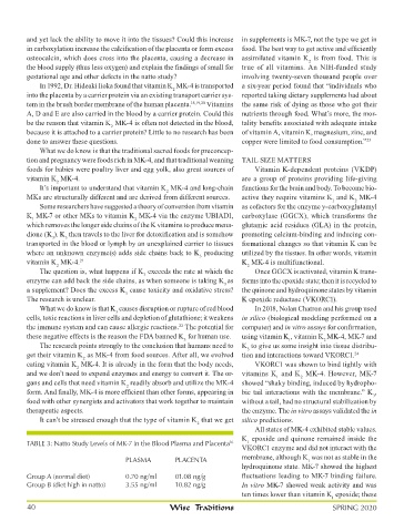
TABLE 3: Natto Study Levels of MK-7 in the Blood Plasma and Placenta 16 1
VKORC1 enzyme and did not interact with the
PLASMA PLACENTA membrane, although K was not as stable in the
1
hydroquinone state. MK-7 showed the highest
Group A (normal diet) 0.70 ng/ml 01.08 ng/g fluctuations leading to MK-7 binding failure.
Group B (diet high in natto) 3.55 ng/ml 10.82 ng/g In vitro MK-7 showed weak activity and was
ten times lower than vitamin K epoxide; these
1
40 Wise Traditions SPRING 2020