
Page 38 - Winter2009
P. 38
In live blood LIVE BLOOD ANALYSIS light delivered through fiber optics. A sterile
Live blood analysis can reveal the presence blood lancet designed for diabetic blood testing
analysis one of certain nutritional deficiencies, such as iron is used to collect a droplet of peripheral blood
can observe deficiency. It also shows the coagulation and from the fingertip, which is immediately placed
the size, clotting processes of the blood, which are related on a microscope slide and then covered with
shape, to the inflammatory biochemical cascade. Fur- a glass cover slip. Oil immersion lenses at the
thermore, the speed of degradation of the blood microscope objective and condenser are used to
variability, removed from the body and observed over time is improve and enhance the images.
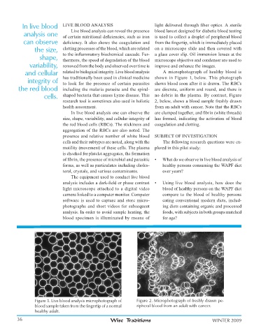
and cellular related to biological integrity. Live blood analysis A microphotograph of healthy blood is
integrity of has traditionally been used in clinical medicine shown in Figure 1, below. This photograph
to look for the presence of certain parasites shows blood soon after it is drawn. The RBCs
the red blood including the malaria parasite and the spiral- are discrete, uniform and round, and there is
cells. shaped bacteria that causes Lyme disease. This no debris in the plasma. By contrast, Figure
research tool is sometimes also used in holistic 2, below, shows a blood sample freshly drawn
health assessment. from an adult with cancer. Note that the RBCs
In live blood analysis one can observe the are clumped together, and fibrin (white threads)
size, shape, variability, and cellular integrity of has formed, indicating the activation of blood
the red blood cells (RBCs). The stickiness and coagulation and clotting.
aggregation of the RBCs are also noted. The
presence and relative number of white blood SUBJECT OF INVESTIGATION
cells and their subtypes are noted, along with the The following research questions were ex-
motility (movement) of these cells. The plasma plored in this pilot study:
is checked for platelet aggregates, the formation
of fibrin, the presence of microbial and parasitic • What do we observe in live blood analysis of
forms, as well as particulates including choles- healthy persons consuming the WAPF diet
terol, crystals, and various contaminants. over years?
The equipment used to conduct live blood
analysis includes a dark-field or phase contrast • Using live blood analysis, how does the
light microscope attached to a digital video blood of healthy persons on the WAPF diet
camera linked to a computer monitor. Computer compare to the blood of healthy persons
software is used to capture and store micro- eating conventional modern diets, includ-
photographs and short videos for subsequent ing diets containing organic and processed
analysis. In order to avoid sample heating, the foods, with subjects in both groups matched
blood specimen is illuminated by means of for age?
Figure 1. Live blood analysis microphotograph of Figure 2. Microphotograph of freshly drawn pe-
blood sample taken from the fingertip of a normal ripheral blood from an adult with cancer.
healthy adult.
36 Wise Traditions WINTER 2009